For many patients, blood transfusion is not an option, whether for religious reasons or safety concerns. The Center for Bloodless Medicine and Surgery team understands and respects this view. We are experts in patient blood management, improving health outcomes and creating peace of mind for those who seek the very best healthcare, without the use of blood or blood products.
Discover the Benefits
There are many benefits to a bloodless approach. Research shows that patients who do not receive blood transfusions recover faster, experience fewer infections and leave the hospital sooner than those who do.How it Works: Bloodless Medicine and Surgery - An Alternative to Blood Transfusion
Bloodless medicine and surgery is an alternative to blood transfusion that among other benefits, has been shown to reduce infections and help patients recover faster. In this video, experts from Johns Hopkins explain the techniques used before, during and after surgery to help patients minimize blood loss and the need to receive donated blood.Bloodless Medicine | Hazel's Story
Hazel Skinner has suffered from chronic illnesses for over 15 years and came to Johns Hopkins from Pennsylvania. Hazel's sister, Dawn, has been to every appointment with her and finds comfort in knowing Hazel's care is a top priority for the Bloodless medicine team. Hazel was admitted to Hopkins and was soon introduced to Dr. Linda Resar, the consultant hematologist for the Bloodless program. Hazel recalls researching the Bloodless program after being told she needed to increase her blood count in able to continue with dialysis. While being sensitive to Hazel’s conscientious refusal to accept blood products, Dr. Resar was able to give her the care she needed to get her hemoglobin back up to healthy levels safely without blood transfusions. After discharge, the Bloodless team continues to follow Hazel. Nurse Coordinator Liz Dackiw calls Hazel regularly to make sure her hemoglobin remains at a healthy level and Hazel continues to be seen by the Bloodless team for regular check-ups throughout the year.
Michael Behe’s introduction of irreducibly complex (IC) molecular machines in Darwin’s Black Box is a gift that keeps on giving. Many readers probably had never heard of cilia or flagella back in 1996. The fact that those machines still make useful illustrations of IC now, even more powerfully than they did 25 years ago, is a strong affirmation of his thesis that IC gives evidence of intelligent design. The bacterial flagellum tends to get more mentions because it is such a cool outboard motor that laypersons can immediately relate to. No less wondrous, though a little more obscure, is the cilium.
Behe updated his description the cilium in his second book, The Edge of Evolution (2007), but research has continued apace. The cilium nails the case for intelligent design more than ever, especially when considering how the organelle is built. Inside those tiny hairlike projections is an advanced transportation system that looks for all the world like a motorized two-way railcar inside a mine shaft!
In Current Biology, Gaia Pigino wrote a “Primer” on Intraflagellar Transport (IFT). It’s called intraflagellar because a cilium is a type of flagellum (Latin for whip), which in the generic sense means a whiplike structure that can move. Both cilia and flagella use the IFT system for construction because both need to transport their building blocks down a shaft from the base to the distal tip. From the railcar’s perspective, the tip would seem a long way away.
There are motile cilia, like the ones that keep our windpipes clean and propel sperm cells, and “primary” cilia, which act as sensory antennae on almost all cells. Accurate construction of cilia is vital. When things go wrong, a host of problems called ciliopathies can result in severe diseases and death. Evolution News has mentioned these briefly in previous years (here, here, and here).
Parts List
Consider first how many players are needed to build a cilium. Pigino’s parts list begins with microtubules in a 9+2 arrangement going up the cilium from base to tip. The two center microtubules are singlets; the outer ring of 9 are in doublet pairs. Riding on those rails are two engines: kinesin-2, which travels from base to tip (anterograde), and dynein-2, which goes from tip back to the base. Kinesin-2 has a head, stalk, hinge and two “feet” (called heads) that walk on the microtubule while carrying a load; the engine contains six protein subunits. Dynein-2 also has a motor, stalk, linker and tail, and is powered by two AAA+ domains that spend ATP for power. Those are the two engine types, and they work in teams along the microtubules.
IFT proteins are numbered, such as IFT8 and IFT176. IFT complexes, such as IFT-A, is composed of six IFT proteins. IFT-B, with 16 IFT proteins is another complex. These ride along the trains to the tip, acting as adaptors for the cargo, which include tubulin proteins, dynein parts, membrane proteins and other IFT proteins.
At the base, a basal body structure called the BBSome forms out of eight BB proteins. It functions as the cargo adaptor for the anterograde train. It authenticates other molecules trying to enter the cilium and moves cargo exiting the retrograde train. Overall, “About 24 different proteins constitute the theoretical minimal functional unit of IFT,” Pigino says, although much needs to be learned.
To address the many fascinating questions that remain about the function and mechanisms of IFT within the cilium and beyond will require the development of new technologies. Fortunately, recent years have seen the introduction of approaches such as cryo-FIB scanning electron microscopy, cryo-correlative light and electron microscopy, and expansion microscopy. The opportunity to combine such approaches with in situ protein tagging for EM, isolation of active IFT complexes, and in vitro reconstitution and reactivation of IFT machinery suggests that IFT studies will continue to yield important insights long into the future. [Emphasis added.]
A precise sequence of amino acids is required for each protein’s function, and the longer the protein, the more improbable that chance could get it right. IFT proteins are large. For instance, BBS1 in the BBSome has 593 amino acid residues; IFT172 (part of the IFT-B complex) has 1,749. The improbability is exacerbated when proteins have to work together. It’s not necessary to belabor the point again, but it’s instructive that Pigino never mentions evolution in her article.
Riding the Train
Moving cargo up and down the cilium takes place in five steps. First, the train assembles at the base. Kinesins line up along a microtubule doublet, their “heads” touching the tracks. Parts of dynein (the return engine) are loaded so as not to touch the tracks, avoiding a “tug-of-war” between the engines. Membrane parts and other cargoes are loaded with the help of IFT-A and IFT-B. Like a well-organized monorail car, the completed train “walks” up the track aided by multiple kinesin-2 engines powered by ATP.
At the tip, the third phase begins. Cargo is unloaded and ferried to the growing cilium (microtubules and membrane). Concurrently, the dynein engines are assembled in an “open configuration as an intermediate state to ensure a controlled activation.” The kinesins are disassembled for transport back to the base. The fourth stage activates the dyneins and starts the train moving, carrying both IFT complexes and waste products to the base. The fifth and final stage unloads the cargo, disassembles the retrograde train and recycles the parts. If you conceive of railcars in a narrow mine shaft carrying tools needed by miners at the far end, and returning the carts with waste products, the analogy seems apt — only the cell’s actions are all automated.
It All Has to Work
Pigino spends much of her Primer discussing ciliopathies: the diseases of broken cilia. When the IFT or engine parts have mutations, or the cilium fails to develop properly, terrible things happen — really terrible things. That’s if the organism (or person) survives at all. Many ciliopathies are not witnessed because the defect causes “major issues during early embryonic development that lead to neonatal death in vertebrates.” She lists 14 known ciliopathies that cause named syndromes, like Bardet-Biedl Syndrome, which causes “rod/cone dystrophy, polydactyly, central obesity, hypogonadism, and kidney dysfunction,” or Retinitis Pigmentosa, which causes blindness. Without getting into the gory details, these ciliar defects can harm the skeleton, eyes, kidneys, brain, or multiple systems in the body at once.
Usually, It Does Work
For the majority of people born with working cilia, here is what they do for us:
Cells need to be able to sense different types of signals, such as chemical and mechanical stimuli, from the extracellular environment in order to properly function. Most eukaryotic cells sense these signals in part through a specialized hair-like organelle, the cilium, that extends from the cell body as a sort of antenna. The signaling and sensory functions of cilia are fundamental during the early stages of embryonic development, when cilia coordinate the establishment of the internal left–right asymmetry that is typical of the vertebrate body. Later, cilia continue to be required for the correct development and function of specific tissues and organs, such as the brain, heart, kidney, liver, and pancreas. Sensory cilia allow us to sense the environment that surrounds us; for instance, we see as a result of the connecting cilia of photoreceptors in our retina, we smell through the sensory cilia at the tips of our olfactory neurons, and we hear thanks to the kinocilia of our sensory hair cells. Motile cilia, which themselves have sensory functions, also work as propeller-like extensions that allow us to breathe because they keep our lungs clean, to reproduce because they propel sperm cells, and even to properly reason because they contribute to the flow of cerebrospinal fluid in our brain ventricles…. Thus, the proper function of cilia is fundamental for human health.
Professor Behe did a wonderful service in introducing these marvelous machines to a wider audience. In 1996, he introduced cilia as examples of irreducible complexity. In 2007, with a decade of new knowledge to draw from, he called cilia examples of “irreducible complexity squared.” Pigina’s Primer on cilia cannot argue with that. If you can breathe, eat, smell, taste, hear, and walk, thank the intelligent designer of cilia that makes these pleasures possible.
Hemoglobin is well known as the molecule that transfers oxygen in blood, but its precursor, heme, is lesser known. Heme is a complex molecule that looks geometrically square, with a single iron atom at the center. The heme family of metalloproteins is responsible for multiple functions in the cell and in the bodies of multicellular organisms, including humans. Our lives depend on heme. When not properly handled, though, it can be dangerous.
What Heme Does
Seven scientists (Galvin Leung et al.) from two UK universities (Leicester, Bristol) explain the significance of heme in their paper, “Unravelling the mechanisms controlling heme supply and demand,” published in PNAS. Their homage to heme is unrestrained, as is their appreciation for how the cell handles this toxic molecule.
Heme is essential for the survival of virtually all living systems and is involved in many fundamental biological processes. It is also implicated as a signaling/regulatory molecule and must be mobilized in response to cellular demands. This presents a complex logistical problem: heme cannot simply diffuse around cells because it is both insoluble and cytotoxic. We show that the cell exhibits exquisite control over release of heme by limiting its availability to one molecule or less within cellular compartments. [Emphasis added.]
Such a description should make a Darwinist shudder. How could such an “exquisite control” system evolve piecemeal? Consider just the making of heme:
Heme is essential for the survival of virtually all living systems — from bacteria, fungi, and yeast, through plants to animals. The family of heme proteins is vast, and heme proteins are responsible for a multitude of functions that are essential for the survival of the cell. To meet the needs of supply and demand for heme in cells, most organisms need to synthesize it. Biosynthesis of the heme cofactor is, therefore, one of the most important metabolic processes in biology; it occurs as an eight-step enzymatic pathway, the last three steps of which occur in the mitochondria.
It takes eight steps to synthesize one heme molecule, and virtually all life needs it — even bacteria, among the simplest of organisms. The other enzymes that construct heme had to already exist before heme could do its job. This is a serious chicken-and-egg problem for the origin of life.
Heme Synthesis
A taste of the complexity of heme synthesis can be had in “Biochemistry, Heme Synthesis,” by Ogun, Joy, and Valentine.
Heme biosynthesis starts in mitochondria with the condensation of succinyl Co-A from the citric acid cycle and an amino acid glycine. They combine to produce a key heme intermediate, 5′-aminolevulinic acid (ALA) in mitochondria catalyzed by the pyridoxal phosphate-requiring (vitamin B6) enzyme, aminolevulinic acid synthase(ALAS). This reaction is the rate-limiting step in the pathway….
That’s just for starters. Those interested in the remaining steps involved in heme synthesis can read four more paragraphs of details like these at the link above. Intermediate forms of the molecule shuttle in and out of a mitochondrion, where special gates control traffic in and out. Multiple other molecules and enzymes, including one metalloprotein containing zinc, are involved in the process.
How could the first cell, by chance, hit on this sequence of steps that would challenge a chemistry grad student? A protocell would have needed to come up with this chemical pathway just to get heme, let alone know what to do with it once it had it. Whoops; it’s toxic, too. How many protocell tryouts died from this essential yet cytotoxic substance before figuring out that heme must be handled with care? Darwinism is dead already — but there’s more.
Heme Supply
The focus of the paper by Leung et al. in PNAS is on how cells distribute heme where it is needed without dying from it.
We suggest an exchange mechanism between protein partners to control supply and demand. Such a mechanism would provide an in-built buffering capacity for heme, enable cells to hoard supplies of heme, and protect the cell against the undesirable effects of heme.
How about that; cells know the law of supply and demand. Where did they learn that? In protocell economics class? They also know how to “hoard supplies” of heme (actually, how to maintain emergency stockpiles). In the recent pandemic, some government officials were aghast to find that emergency stockpiles of PPE (personal protective equipment), required by law, had been raided or not maintained. It led to serious shortages and drastic efforts to refill stockpiles, while patients were dying and healthcare workers were exposed to the virus without protection. Cells do not make such mistakes.
Heme Distribution
Scientists have known all about heme and its functions for decades, but few have investigated how cells distribute it where needed. This is important to know, Leung et al. explain, because “Deficiencies or excesses in cellular heme concentration also have widespread implications in health and disease” such as aging, cardiovascular disease, inflammation, and immune response. Accordingly, “there is a need to understand the logistics of heme supply and demand.”
A cell cannot maintain a “pool” of heme to draw from, as once thought, because heme is a “nuisance” to cells. It tends form free radicals, which are dangerous, and though hydrophobic, it dimerizes in solution, making it unsuitable for delivery to proteins that only need one heme molecule per binding site.
A free molecule of heme can therefore only exist transiently, and if a large reserve of heme is present, the heme molecules would presumably need to be exchanged rapidly between binding partners to remain solubilized, in the same way that heme is solubilized within the interior of other well-known heme proteins (e.g., hemoglobin).
The team constructed a molecular heme sensor that glows when bound to heme. In this way, they could watch the “exquisite control” system in action.
A longstanding question has been to establish the mechanisms that control the supply and demand for cellular heme. … we have developed a heme sensor … that can respond to heme availability…. The results demonstrate that concentrations are typically limited to one molecule or less within cellular compartments. These miniscule amounts of free heme are consistent with a system that sequesters the heme and is able to buffer changes in heme availability while retaining the capability to mobilize heme when and where it is needed. … This exquisite control, in which heme is made available for transfer one molecule at a time, protects the cellagainst the toxic effect of excess heme and offers a simple mechanism for heme-dependent regulation in single-molecule steps.
In effect, the cell maintains “an exchangeable (buffered) heme reservoir” that solves the availability problem while simultaneously protecting the cell from heme’s toxic effects. Free heme (the risky kind) was detected only in “a minute fraction of the entire amount of heme present in the cell” and were most likely short-lived in the process of binding to proteins.
Our experiments are thus consistent with the idea that there is a population of the total heme complement that is bound more weakly and therefore reversibly to heme-binding partner proteins or to other molecules (which might include free amino acids) that can buffer against changes in the heme concentration. … These heme molecules that are weakly bound to buffer molecules, along with the miniscule population of free heme, would constitute a body of exchangeable heme in the cell.
In their model, the buffered heme, attached to its partner, passes quickly to the enzyme or protein needing it, something like a quick pass of the ball from one player to another in basketball or football. In the cell’s game, though, there are millions of balls with millions of players passing the heme balls to the players who need it. Because the free energy of the acceptor is at a lower level, the heme is readily transferred to the acceptor, leaving the partner ready to pick up another heme. At any given time, the cell can be aware of the concentration of available heme by sensing the concentration of heme-binding partners, and supply more as the demand increases.
This exquisite control also provides a mechanism for heme-dependent signaling and regulation, as heme can be supplied discretely, leading to the switching on of proteins in single-molecule steps.
If Darwinism had been essential to their work, they surely would have mentioned it. Instead, they found a mechanism that appears (gasp! Can they say this?) designed for a purpose —
We see clear advantages of such an exchange mechanism between protein partners, designed for the purpose of managing heme supply and demand.
Overkill
To nail the case for design, consider the level of exquisite control in the next hierarchical level up. The human body makes around 250 billion red blood cells per day, and each RBC contains 270 million hemoglobin molecules, each constructed with 4 heme groups. That multiplies out to 27 billion trillion hemes per day!
It’s amazing enough that each cell in the body orchestrates its synthesis and availability of heme. On top of that, the whole body, too, regulates the number of hemoglobin molecules and red blood cells that carry another cytotoxic substance — oxygen — from our lungs to each cell in a safe, regulated, exquisitely controlled manner. Every red-blooded person should take this to heart: we would be walking packages of explosives if it were not for mechanisms “designed for the purpose” of using energy safely for life, love, and transcendent meaning.
As I explained in a post yesterday, a TEDx talk from January of this year by MIT bioengineer Erika DeBenedictis, “It’s Time for Intelligent Design,” argues that biology lacks any “intelligent design,” and thus living systems are “imperfect” with “gigantic mistakes,” and we should “play God” in order to “make biology better.” So what are some flaws in biology that we can make “better”? According to Dr. DeBenedictis, “one of the big limitations of biology are the basic building blocks themselves.” Emily Reeves has already addressed Dr. DeBenedictis’s comments about the optimality of amino acids in the genetic code, noting that the set of amino acids used by life is highly optimal. But DeBenedictis goes further and says that the very makeup of the building blocks of life need a major redesign.
“Kind of Boring”
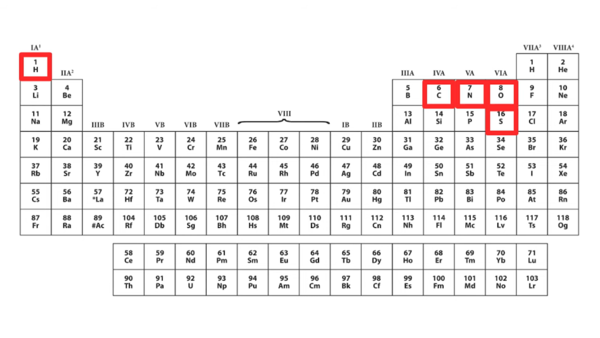
“If we look at the amino acids,” she states, “chemically speaking, they’re actually kind of boring” and “one very easy way to see this is to look at them on a periodic table.” She then shows a periodic table that highlights five chemical elements — hydrogen, carbon, nitrogen, oxygen, and sulfur — she says “Out of all this good stuff, only five chemical elements appear in proteins.” Here’s the slide from the talk:
If we are to take Dr. DeBenedictis at her word, then “only five chemical elements appear in proteins,” namely those highlighted in her chart: hydrogen, carbon, nitrogen, oxygen, and sulfur. She further says she wants to create new building blocks and says, “maybe we could get some heavy metals up here.” The message is that proteins only use five elements and that proteins and enzymes essentially ignore the rest of the period table — more evidence of what she calls “imperfect design.”
Dr. DeBenedictis is right that amino acids themselves don’t contain metals. But is it correct for her to state that “only five chemical elements appear in proteins,” suggesting that the rest of the periodic table is absent from protein complexes and not involved in enzyme chemistry? Is it true that proteins don’t make use of metals? No, that is not correct.
Metals, Metals, Everywhere…
Enzymes are proteins that catalyze reactions, and Voet and Voet’s standard textbook Biochemistry states “Nearly one-third of all known enzymes require the presence of metal ions for catalytic activity.” (Voet and Voet 2004, p. 501). The textbook continues:
There are two classes of metal ion-requiring enzymes that are distinguished by the strengths of their ion-protein interactions:
1. Metalloenzymes contain tightly bound metal ions, most commonly transition metal ions such as Fe2+, Fe3+, Cu2+, Zn2+, Mn2+, or Co3+.
2. Metal-activated enzymes loosely bind metal ions from solution, usually the alkali and alkaline earth metal ions Na+, K+, Mg2+, or Ca2+.
Metal ions participate in the catalytic process in three major ways:
1. By binding to substrates so as to orient them properly for reaction.
2. By mediating oxidation-reduction reactions through reversible changes in the metal ion’s oxidation state.
3. By electrostatically stabilizing or shielding negative charges.
Metal ions are constituents of many metalloproteins, in which they have either catalytic (metalloenzymes) or structural functions. … Metals are important for the biological activity of proteins and their removal or the replacement of one metal by another is often accompanied by a loss of or reduction in the biological activity of the protein. Metal ions in proteins can act as structure promoters or can take part in enzymatic reactions. Divalent metal cations such as Zn2+, Mg2+, Cu2+ and Ca2+ are often associated with the catalytic or regulatory activities of proteins that constitute some of the fundamental chemical life processes. For example, Mg in chlorophyll is important for photosynthesis, Cu (together with Fe) has a role in oxygen-carrying proteins and Zn, Mg and Ca can serve as Lewis acids. (emphasis added, internal citations omitted)
A 2011 entry on metalloenzymes in Springer’s Encyclopedia of Geobiology likewise explains that up to a third of known enzymes — i.e., an important type of protein that fosters chemical reactions in cells — use metals as cofactors:
Metalloenzymes are enzyme proteins containing metal ions (metal cofactors), which are directly bound to the protein or to enzyme-bound nonprotein components (prosthetic groups). About one-third of all enzymes known so far are metalloenzymes… metalloenzymes are found in all enzymes families…
The encyclopedia entry goes on to explain:
Besides enzymes, other metalloproteins are involved in non-enzyme electron transfer reactions (cytochromes), may act as storage (e.g., ferritin for iron) or transport proteins (e.g., transferrin for iron). In the latter groups of proteins, the metal storage is reversible and the metal is a temporary component. Also, ribozymes, i.e., RNA molecules with enzyme function may contain structurally and/or functionally important metal ions … and may be therefore termed as metalloenzymes in a broader sense.
Later the entry lists a table summarizing the functions of various metals in enzymes — here’s a condensed version of that table:
Metallic Element
Roles Played in Enzymes
Potassium
Protein stabilization
Calcium
Protein stabilization
Magnesium
Protein stabilization
Iron
Oxygen transport, storage and/or activation; electron transport; superoxide breakdown
Manganese
Oxygen release; peroxide and superoxide breakdown
Zinc
Protein stabilization; hydrolytic cleavage
Copper
Oxygen transport, storage and/or activation; electron transport; superoxide breakdown
Molybdenum
Oxygen transfer; N2 activation
Cobalt
Free radical reactions; nucleophile
Going through the entry, it becomes evident that the variety and number of uses of metals in enzymes and proteins are almost endless. Examples listed include:
“Mononuclear iron proteins are oxidoreductases (e.g., aromatic amino acid hydroxylases, aromatic ring cleavage dioxygenases, Fe-superoxide dismutase, lipoxygenases, nitrile hydratase, and Rieske oxygenases), or involved in electron transfer (desulforedoxins, rubredoxins, andphotosynthetic reaction centers).”
“Hemoproteins are characterized by an iron porphyrin as prosthetic group” and include “oxidoreductases (catalases, peroxidases), electron transfer proteins (cytochromes), or oxygen transport and storage proteins (globins).”
“Iron–sulfur proteins are characterized by the presence of iron-sulfur clusters containing sulfide-linked di-, tri-, and tetra iron centers in variable oxidation states” and include “ferredoxins, NADH hydrogenases, dehydrogenases, cytochrome creductases, nitrogenases, and other proteins.”
“Copper proteins are involved in oxygen transport or activation processes and electron transport…” and include “ascorbate oxidase, ceruloplasmin, laccase, nitritereductase, auracyanin, and azurin …”
Heavy Metals
Dr. DeBenedictis’s precise statement in her talk says that we should add “heavy metals” to life. It’s not exactly clear how she would define “heavy metals,” but her slide (see above) and language seem to suggest that proteins don’t use any metals at all. While she is likely trying to simplify things to make a point, it is incorrect to say that proteins don’t use heavy metals. For example, a 2020 paper in Scientific Reports states that many “heavy metals” are “bioavailable,” and these include zinc (Zn), copper (Cu), and nickel (Ni). Multiple sources explain how widely used these heavy metals are in plants.
Zinc: A 2016 study notes how important zinc is in plants:
Zinc (Zn) is an essential micronutrient for plant growth and development. It is also a crucial element for survival of most of the organisms including humans. Zn acts as cofactor of more than 300 proteins, among which majority are zinc finger proteins, RNA polymerases and DNA polymerases. It is the only metal present in all six enzyme classes (oxidoreductase, transferase, hydrolases, lyases, isomerases and ligases). Being a part of structural or catalytic units, Zn regulates the activities, conformational stabilization and folding of various proteins (enzymes). In addition to this, role of Zn in membrane integrity and stabilization, alleviation of oxidative stress — and as intracellular second messenger has also been reported. Zn is involved in a number of plant physiological processes such as hormone regulation (e.g. tryptophan synthesis, a precursor of IAA), signal transduction via mitogen activated protein kinases, repair processes of PS II complex during photoinhibition and maintenance of CO2 concentration in mesophyll. Peck and McDonald (2010) — confirms the participation of Zn in regulation of Rubisco activity along with alleviation from adverse effects of heat stress in wheat. Thus, Zn essentiality for plant system is illustrated from literature cited above.” [Internal citations removed.]
Zinc activates enzymes that are responsible for the synthesis of certain proteins. It is used in the formation of chlorophyll and some carbohydrates, conversion of starches to sugars and its presence in plant tissue helps the plant to withstand cold temperatures. Zinc is essential in the formation of auxins, which help with growth regulation and stem elongation.
Zinc plays similar roles in animal biochemistry.
Copper: A 2018 paper notes that “copper is an essential element for proper growth and development of plants. Copper in plants is functioning as a catalyst in respiration and photosynthesis. It is vital element for the creation of lignin in plant cell walls and in the case of enzymes responsible for protein synthesis. It also influences the disease resistance and reproduction.” The horticulture site likewise explains the role of copper in plants:
Copper activates some enzymes in plants which are involved in lignin synthesis and it is essential in several enzyme systems. It is also required in the process of photosynthesis, is essential in plant respiration and assists in plant metabolism of carbohydrates and proteins. Copper also serves to intensify flavor and color in vegetables and color in flowers.
Nickel: As for nickel, the same horticulture website explains:
Nickel is a component of some plant enzymes, most notably urease, which metabolizes urea nitrogen into useable ammonia within the plant. Without nickel, toxic levels of urea can accumulate within the tissue forming necrotic legions on the leaf tips. In this case, nickel deficiency causes urea toxicity. Nickel is also used as a catalyst in enzymes used to help legumes fix nitrogen. There is evidence that nickel helps with disease tolerance in plants, although it is still unclear how this happens.
Ideas Have Consequences
I could go on, citing review papers, research papers, biochemistry textbooks, and other authorities explaining how important metals are to the basic functions of numerous proteins. At the very least, it seems an oversight to criticize biology for not using “heavy metals” and yet fail to acknowledge any of the numerous instances where metals are used to fulfill core functions in enzymes and other proteins. To show a full periodic table that highlights only hydrogen, carbon, nitrogen, oxygen, and sulfur, and then to say “Out of all this good stuff, only five chemical elements appear in proteins,” severely underrepresents the degree to which metals are used in biology.
I infer no bad faith or ignorance on Dr. DeBenedictis’s part. Although her Covid-lockdown-era TEDx talk was recorded privately, with opportunities for multiple takes to get things right, I believe what she probably meant was that amino acids don’t use metals, and it was a simple misstatement. But there’s still a lesson here.
Ideas have consequences. When we adopt a materialistic perspective which assumes that biology has no intelligent design, and instead contains fundamental flaws or “gigantic mistakes,” we’ll be quicker to assume that features of biological systems exist for no good reason. This discourages us from investigating why things are the way they are. We’ll be more likely to miss good design features in biology, which is exactly what I think has happened here.
Materialism’s blind spots often exist right where important biological features are found. We’ve certainly seen this blind spot in past thinking about “junk DNA,” where a standard evolutionary framework discouraged research into the functions of this DNA. Today, we know “junk DNA” has key biological functions. But materialistic thinking doesn’t encourage us to understand the design principles that underly biology.
Intelligent design is an idea that has consequences, too. And when it comes to understanding biology, ID is a paradigm that can lead to fruitful research predictions. Those predictions start with the suspicion that there’s probably a good reason why things are the way they are, and task is up to us to figure out what those reasons are.
I have been discussing Darwin’s “abominable mystery” — the abrupt origin of flowering plants (see here and here). I noted that two new studies claim progress in solving this problem. This year a new article by Silvestro et al. (2021), “Fossil data support a pre-Cretaceous origin of flowering plants,” was published in the prestigious journal Nature Ecology & Evolution. It claimed that the fossil record proves that flowering plants already existed in the Jurassic period or even earlier. Sounds like they finally discovered the elusive Jurassic ancestors of flowering plants!? Indeed, the popular press headlined, “Flowering plants may be 100 million years older than we thought” (Sawal 2021) and “Study: First Flowering Plants Appeared in Jurassic Period or Even Earlier” (SciNews 2021). The official press release announced, “New study unravels Darwin’s ‘abominable mystery’ surrounding origin of flowering plants” (University of Bristol 2021).
Did It Really?
In spite of the hype, this study is highly problematic. You do not have to take my word for that, because even some mainstream evolutionists agree, such as Patrick Herendeen from the Chicago Botanic Garden in a skeptical comment quoted in New Scientist (Sawal 2021). Herendeen is skeptical of the findings “because missing fossil data could affect the results.” Missing fossil data? Sounds suspicious, so, what did the new study really discover to justify the grand claims? Silvestro et al. (2021) looked at the statistical distribution of the oldest fossil representatives for modern angiosperm families only, which are exclusively Tertiary and Cretaceous in age. Similar to many molecular clock studies (Bell et al. 2005, 2010, Smith et al. 2010), which totally contradict the fossil record (Coiro et al. 2019), and similar to some other studies (Li et al. 2019), they then extrapolated the history of these groups back into the Jurassic with a so-called ghost lineage, even though there are no Jurassic fossils. You heard that right: They did not find any Jurassic fossils of flowering plants, not a single one! To explain away the conflicting evidence, the authors had to invoke a completely ad hoc hypothesis that was pondered by Charles Darwin in his 1879 letter to Hooker, namely that angiosperms evolved slowly as a rare and small group in a remote and hidden region (Barba-Montoya et al. 2018, Sgorbati et al. 2018), which therefore evaded the fossil record. But if we consider the numerous fossil localities around the globe with thousands of Jurassic plant fossils, but no angiosperms among them, this cheap cop-out is simply not plausible and not warranted by any positive evidence. It is a desperate attempt to shield a cherished theory against empirical falsification, and that is definitely not how good science should be done.
An Even More Fundamental Problem
But there is an even more fundamental problem with this study. The authors’ claim is based on two question-begging assumptions: common descent (which I would even grant), and a gradual, slow neo-Darwinian mechanism of evolution. Only based on these two assumptions can they postulate and extrapolate a ghost lineage back into the Jurassic. The actual fossil record clearly contradicts their prediction, and thus suggests that at least one of their two hidden assumptions is wrong. In my view this is clearly the neo-Darwinian unguided process, which is also undermined by many other lines of evidence, such as the waiting time problem or the combinatorial search space problem. This is why neo-Darwinism is indeed increasingly questioned and rejected by leading theoretical biologists (Nelson & Klinghoffer 2016), and not just by us Darwin critics and intelligent design proponents.
The study by Silvestro et al. (2021) does not unravel but rather confirms Darwin’s “abominable mystery,” because it shows that Darwin’s intuition was correct: if the evolution of angiosperms before the Cretaceous was at the same rate/mode/tempo as the evolution of angiosperms after the Cretaceous, then there should be Jurassic angiosperm fossils. Given that Jurassic angiosperm fossils have not been found, this study provides new confirmation of the abominable mystery, not its solution. That is not just my humble opinion, but has also been expressed in two tweets by Richard Buggs (see Twitter, here and here).
Next, “Darwin’s ‘Abominable Mystery’: Mesozoic Cupules Come to the Rescue?”