Search This Blog

Friday 18 June 2021

Playing out of their league?


Do Proteins Lack Metals, Reflecting Poor Design?

Casey Luskin

As I explained in a post yesterday, a TEDx talk from January of this year by MIT bioengineer Erika DeBenedictis, “It’s Time for Intelligent Design,” argues that biology lacks any “intelligent design,” and thus living systems are “imperfect” with “gigantic mistakes,” and we should “play God” in order to “make biology better.” So what are some flaws in biology that we can make “better”? According to Dr. DeBenedictis, “one of the big limitations of biology are the basic building blocks themselves.” Emily Reeves has already addressed Dr. DeBenedictis’s comments about the optimality of amino acids in the genetic code, noting that the set of amino acids used by life is highly optimal. But DeBenedictis goes further and says that the very makeup of the building blocks of life need a major redesign.

“Kind of Boring”

“If we look at the amino acids,” she states, “chemically speaking, they’re actually kind of boring” and “one very easy way to see this is to look at them on a periodic table.” She then shows a periodic table that highlights five chemical elements — hydrogen, carbon, nitrogen, oxygen, and sulfur — she says “Out of all this good stuff, only five chemical elements appear in proteins.” Here’s the slide from the talk:

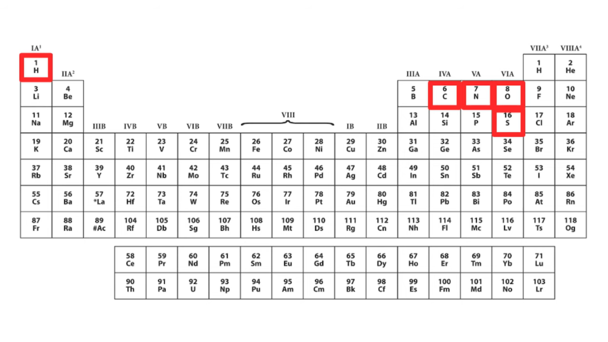
If we are to take Dr. DeBenedictis at her word, then “only five chemical elements appear in proteins,” namely those highlighted in her chart: hydrogen, carbon, nitrogen, oxygen, and sulfur. She further says she wants to create new building blocks and says, “maybe we could get some heavy metals up here.” The message is that proteins only use five elements and that proteins and enzymes essentially ignore the rest of the period table — more evidence of what she calls “imperfect design.”

Dr. DeBenedictis is right that amino acids themselves don’t contain metals. But is it correct for her to state that “only five chemical elements appear in proteins,” suggesting that the rest of the periodic table is absent from protein complexes and not involved in enzyme chemistry? Is it true that proteins don’t make use of metals? No, that is not correct. 

Metals, Metals, Everywhere…

Enzymes are proteins that catalyze reactions, and Voet and Voet’s standard textbook Biochemistry states “Nearly one-third of all known enzymes require the presence of metal ions for catalytic activity.” (Voet and Voet 2004, p. 501). The textbook continues:

There are two classes of metal ion-requiring enzymes that are distinguished by the strengths of their ion-protein interactions:

1. Metalloenzymes contain tightly bound metal ions, most commonly transition metal ions such as Fe2+, Fe3+, Cu2+, Zn2+, Mn2+, or Co3+.

2. Metal-activated enzymes loosely bind metal ions from solution, usually the alkali and alkaline earth metal ions Na+, K+, Mg2+, or Ca2+.

Metal ions participate in the catalytic process in three major ways:

1. By binding to substrates so as to orient them properly for reaction.

2. By mediating oxidation-reduction reactions through reversible changes in the metal ion’s oxidation state.

3. By electrostatically stabilizing or shielding negative charges.

VOET AND VOET’S, BIOCHEMISTRY, 2004, P. 501

A 2008 review in Acta Crystallographica Section D, titled “Metals in proteins: correlation between the metal-ion type, coordination number and the amino-acid residues involved in the coordination,” similarly notes how prevalent and important metal usage is in proteins:

Metal ions are constituents of many metalloproteins, in which they have either catalytic (metalloenzymes) or structural functions. … Metals are important for the biological activity of proteins and their removal or the replacement of one metal by another is often accompanied by a loss of or reduction in the biological activity of the protein. Metal ions in proteins can act as structure promoters or can take part in enzymatic reactions. Divalent metal cations such as Zn2+, Mg2+, Cu2+ and Ca2+ are often associated with the catalytic or regulatory activities of proteins that constitute some of the fundamental chemical life processes. For example, Mg in chlorophyll is important for photosynthesis, Cu (together with Fe) has a role in oxygen-carrying proteins and Zn, Mg and Ca can serve as Lewis acids. (emphasis added, internal citations omitted)

A 2011 entry on metalloenzymes in Springer’s Encyclopedia of Geobiology likewise explains that up to a third of known enzymes — i.e., an important type of protein that fosters chemical reactions in cells — use metals as cofactors:

Metalloenzymes are enzyme proteins containing metal ions (metal cofactors), which are directly bound to the protein or to enzyme-bound nonprotein components (prosthetic groups). About one-third of all enzymes known so far are metalloenzymes… metalloenzymes are found in all enzymes families…

The encyclopedia entry goes on to explain:

Besides enzymes, other metalloproteins are involved in non-enzyme electron transfer reactions (cytochromes), may act as storage (e.g., ferritin for iron) or transport proteins (e.g., transferrin for iron). In the latter groups of proteins, the metal storage is reversible and the metal is a temporary component. Also, ribozymes, i.e., RNA molecules with enzyme function may contain structurally and/or functionally important metal ions … and may be therefore termed as metalloenzymes in a broader sense.

Later the entry lists a table summarizing the functions of various metals in enzymes — here’s a condensed version of that table:

Metallic ElementRoles Played in Enzymes
PotassiumProtein stabilization
CalciumProtein stabilization
MagnesiumProtein stabilization
IronOxygen transport, storage and/or activation; electron transport; superoxide breakdown
ManganeseOxygen release; peroxide and superoxide breakdown
ZincProtein stabilization; hydrolytic cleavage
CopperOxygen transport, storage and/or activation; electron transport; superoxide breakdown
MolybdenumOxygen transfer; N2 activation
CobaltFree radical reactions; nucleophile

Going through the entry, it becomes evident that the variety and number of uses of metals in enzymes and proteins are almost endless. Examples listed include:

  • “Mononuclear iron proteins are oxidoreductases (e.g., aromatic amino acid hydroxylases, aromatic ring cleavage dioxygenases, Fe-superoxide dismutase, lipoxygenases, nitrile hydratase, and Rieske oxygenases), or involved in electron transfer (desulforedoxins, rubredoxins, andphotosynthetic reaction centers).”
  • “Hemoproteins are characterized by an iron porphyrin as prosthetic group” and include “oxidoreductases (catalases, peroxidases), electron transfer proteins (cytochromes), or oxygen transport and storage proteins (globins).”
  • “Iron–sulfur proteins are characterized by the presence of iron-sulfur clusters containing sulfide-linked di-, tri-, and tetra iron centers in variable oxidation states” and include “ferredoxins, NADH hydrogenases, dehydrogenases, cytochrome creductases, nitrogenases, and other proteins.”
  • “Copper proteins are involved in oxygen transport or activation processes and electron transport…” and include “ascorbate oxidase, ceruloplasmin, laccase, nitritereductase, auracyanin, and azurin …”

Heavy Metals

Dr. DeBenedictis’s precise statement in her talk says that we should add “heavy metals” to life. It’s not exactly clear how she would define “heavy metals,” but her slide (see above) and language seem to suggest that proteins don’t use any metals at all. While she is likely trying to simplify things to make a point, it is incorrect to say that proteins don’t use heavy metals. For example, a 2020 paper in Scientific Reports states that many “heavy metals” are “bioavailable,” and these include zinc (Zn), copper (Cu), and nickel (Ni). Multiple sources explain how widely used these heavy metals are in plants.

Zinc: 2016 study notes how important zinc is in plants:

Zinc (Zn) is an essential micronutrient for plant growth and development. It is also a crucial element for survival of most of the organisms including humans. Zn acts as cofactor of more than 300 proteins, among which majority are zinc finger proteins, RNA polymerases and DNA polymerases. It is the only metal present in all six enzyme classes (oxidoreductase, transferase, hydrolases, lyases, isomerases and ligases). Being a part of structural or catalytic units, Zn regulates the activities, conformational stabilization and folding of various proteins (enzymes). In addition to this, role of Zn in membrane integrity and stabilization, alleviation of oxidative stress — and as intracellular second messenger has also been reported. Zn is involved in a number of plant physiological processes such as hormone regulation (e.g. tryptophan synthesis, a precursor of IAA), signal transduction via mitogen activated protein kinases, repair processes of PS II complex during photoinhibition and maintenance of CO2 concentration in mesophyll. Peck and McDonald (2010) — confirms the participation of Zn in regulation of Rubisco activity along with alleviation from adverse effects of heat stress in wheat. Thus, Zn essentiality for plant system is illustrated from literature cited above.” [Internal citations removed.]

According to a horticulture website:

Zinc activates enzymes that are responsible for the synthesis of certain proteins. It is used in the formation of chlorophyll and some carbohydrates, conversion of starches to sugars and its presence in plant tissue helps the plant to withstand cold temperatures. Zinc is essential in the formation of auxins, which help with growth regulation and stem elongation.

Zinc plays similar roles in animal biochemistry. 

Copper: 2018 paper notes that “copper is an essential element for proper growth and development of plants. Copper in plants is functioning as a catalyst in respiration and photosynthesis. It is vital element for the creation of lignin in plant cell walls and in the case of enzymes responsible for protein synthesis. It also influences the disease resistance and reproduction.” The horticulture site likewise explains the role of copper in plants:

Copper activates some enzymes in plants which are involved in lignin synthesis and it is essential in several enzyme systems. It is also required in the process of photosynthesis, is essential in plant respiration and assists in plant metabolism of carbohydrates and proteins. Copper also serves to intensify flavor and color in vegetables and color in flowers.

Nickel: As for nickel, the same horticulture website explains:

Nickel is a component of some plant enzymes, most notably urease, which metabolizes urea nitrogen into useable ammonia within the plant. Without nickel, toxic levels of urea can accumulate within the tissue forming necrotic legions on the leaf tips. In this case, nickel deficiency causes urea toxicity. Nickel is also used as a catalyst in enzymes used to help legumes fix nitrogen. There is evidence that nickel helps with disease tolerance in plants, although it is still unclear how this happens.

Ideas Have Consequences

I could go on, citing review papers, research papers, biochemistry textbooks, and other authorities explaining how important metals are to the basic functions of numerous proteins. At the very least, it seems an oversight to criticize biology for not using “heavy metals” and yet fail to acknowledge any of the numerous instances where metals are used to fulfill core functions in enzymes and other proteins. To show a full periodic table that highlights only hydrogen, carbon, nitrogen, oxygen, and sulfur, and then to say “Out of all this good stuff, only five chemical elements appear in proteins,” severely underrepresents the degree to which metals are used in biology. 

I infer no bad faith or ignorance on Dr. DeBenedictis’s part. Although her Covid-lockdown-era TEDx talk was recorded privately, with opportunities for multiple takes to get things right, I believe what she probably meant was that amino acids don’t use metals, and it was a simple misstatement. But there’s still a lesson here. 

Ideas have consequences. When we adopt a materialistic perspective which assumes that biology has no intelligent design, and instead contains fundamental flaws or “gigantic mistakes,” we’ll be quicker to assume that features of biological systems exist for no good reason. This discourages us from investigating why things are the way they are. We’ll be more likely to miss good design features in biology, which is exactly what I think has happened here. 

Materialism’s blind spots often exist right where important biological features are found. We’ve certainly seen this blind spot in past thinking about “junk DNA,” where a standard evolutionary framework discouraged research into the functions of this DNA. Today, we know “junk DNA” has key biological functions. But materialistic thinking doesn’t encourage us to understand the design principles that underly biology. 

Intelligent design is an idea that has consequences, too. And when it comes to understanding biology, ID is a paradigm that can lead to fruitful research predictions. Those predictions start with the suspicion that there’s probably a good reason why things are the way they are, and task is up to us to figure out what those reasons are. 

Monday 14 June 2021

Flower power vs. Darwin round 2.


Darwin’s “Abominable Mystery”: Jurassic Flowering Plants After All?

Günter Bechly

I have been discussing Darwin’s “abominable mystery” — the abrupt origin of flowering plants (see here and here). I noted that two new studies claim progress in solving this problem. This year a new article by Silvestro et al. (2021), “Fossil data support a pre-Cretaceous origin of flowering plants,” was published in the prestigious journal Nature Ecology & Evolution. It claimed that the fossil record proves that flowering plants already existed in the Jurassic period or even earlier. Sounds like they finally discovered the elusive Jurassic ancestors of flowering plants!? Indeed, the popular press headlined, “Flowering plants may be 100 million years older than we thought” (Sawal 2021) and “Study: First Flowering Plants Appeared in Jurassic Period or Even Earlier” (SciNews 2021). The official press release announced, “New study unravels Darwin’s ‘abominable mystery’ surrounding origin of flowering plants” (University of Bristol 2021).

Did It Really?

In spite of the hype, this study is highly problematic. You do not have to take my word for that, because even some mainstream evolutionists agree, such as Patrick Herendeen from the Chicago Botanic Garden in a skeptical comment quoted in New Scientist (Sawal 2021). Herendeen is skeptical of the findings “because missing fossil data could affect the results.” Missing fossil data? Sounds suspicious, so, what did the new study really discover to justify the grand claims? Silvestro et al. (2021) looked at the statistical distribution of the oldest fossil representatives for modern angiosperm families only, which are exclusively Tertiary and Cretaceous in age. Similar to many molecular clock studies (Bell et al. 20052010Smith et al. 2010), which totally contradict the fossil record (Coiro et al. 2019), and similar to some other studies (Li et al. 2019), they then extrapolated the history of these groups back into the Jurassic with a so-called ghost lineage, even though there are no Jurassic fossils. You heard that right: They did not find any Jurassic fossils of flowering plants, not a single one! To explain away the conflicting evidence, the authors had to invoke a completely ad hoc hypothesis that was pondered by Charles Darwin in his 1879 letter to Hooker, namely that angiosperms evolved slowly as a rare and small group in a remote and hidden region (Barba-Montoya et al. 2018Sgorbati et al. 2018), which therefore evaded the fossil record. But if we consider the numerous fossil localities around the globe with thousands of Jurassic plant fossils, but no angiosperms among them, this cheap cop-out is simply not plausible and not warranted by any positive evidence. It is a desperate attempt to shield a cherished theory against empirical falsification, and that is definitely not how good science should be done.

An Even More Fundamental Problem

But there is an even more fundamental problem with this study. The authors’ claim is based on two question-begging assumptions: common descent (which I would even grant), and a gradual, slow neo-Darwinian mechanism of evolution. Only based on these two assumptions can they postulate and extrapolate a ghost lineage back into the Jurassic. The actual fossil record clearly contradicts their prediction, and thus suggests that at least one of their two hidden assumptions is wrong. In my view this is clearly the neo-Darwinian unguided process, which is also undermined by many other lines of evidence, such as the waiting time problem or the combinatorial search space problem. This is why neo-Darwinism is indeed increasingly questioned and rejected by leading theoretical biologists (Nelson & Klinghoffer 2016), and not just by us Darwin critics and intelligent design proponents.

The study by Silvestro et al. (2021) does not unravel but rather confirms Darwin’s “abominable mystery,” because it shows that Darwin’s intuition was correct: if the evolution of angiosperms before the Cretaceous was at the same rate/mode/tempo as the evolution of angiosperms after the Cretaceous, then there should be Jurassic angiosperm fossils. Given that Jurassic angiosperm fossils have not been found, this study provides new confirmation of the abominable mystery, not its solution. That is not just my humble opinion, but has also been expressed in two tweets by Richard Buggs (see Twitter, here and here).

Next, “Darwin’s ‘Abominable Mystery’: Mesozoic Cupules Come to the Rescue?”


Sunday 13 June 2021

Darwinism's ministry of truth is at it again.


Ixnay on the Ambriancay Plosionexhay

Paul Nelson

Shhh — don’t say it. Don’t say, you know, that. The geological event, well, not “event,” as in something that happened about 541 million years ago…we don’t talk that way anymore.

Because people may get the wrong idea. And we don’t want them to have those bad thoughts and wrong ideas. Changing the names of things helps people to think correctly. Right?

A dozen authors at Michigan State University have proposed that the paleontological term “Cambrian Explosion” be laid “to rest for any use other than historical reference.” The problem, as they see it, is the tendency of “anti-science” readers to misappropriate “Cambrian Explosion,” to suggest that evolution somehow fails to explain the appearance of nearly all the animal phyla without fossil antecedents of equal complexity. As they argue:

Certainly, biodiversification at the beginning of the Cambrian was unique (Erwin et al., 1987) — all those new body plans — but no evolutionary rules were broken, nor is there mystery or discipline-dividing controversy, as is claimed by anti-science concerns who seize on the term “explosion.”

Their alternative? “Great Cambrian Biodiversification,” or GCB for short.

GCB, alas, may have a short life of its own, because the geological data haven’t changed. It isn’t the name of the paleontological pattern of sudden appearance, at the base of the Cambrian, that matters at the end of the day. Call that pattern “Wayne Q. Jones” or “cured sausage” or “Fossils-a-plenty,” the evidence in the rocks is what it is. When students find out about the fossil evidence, “GCB” will be just as bothersome to certain evolutionary sensibilities as “Cambrian Explosion” appears to be today. 

GCB = Cambrian Explosion = a pattern that puzzled Charles Darwin over 160 years ago. Change the name, and the puzzle lives on.


Saturday 12 June 2021

Flower power vs. Darwin

Darwin’s “Abominable Mystery”: Still Alive and Kicking

Günter Beckly

 a podcast for ID the Future (Bechly 2021), I discussed some recent articles about Darwin’s “abominable mystery” of the abrupt origin of flowering plants in the Cretaceous period, and showed that this mystery is not only still unresolved, but has even become much more mysterious since Darwin’s time.

Now, two new studies claim progress in solving this mystery. But before we look into these new studies, let’s first explore some background context on this fascinating issue.

“As Fully Formed as Aphrodite”

The crucial issue is a remarkable phenomenon in the fossil record, namely the sudden appearance of fossils of flowering plants in the Early Cretaceous age without any of the required precursors recorded from the older Jurassic layers. In the mid Cretaceous there is already a large diversity of at least 50 different families of flowering plants, as if they came out of nowhere or were planted there by a creator. This fact is beautifully captured in a comment by Oskin (2015) about the highly controversial discovery of a Jurassic fossil flower:

Then, about 125 million years ago, angiosperms and their flowers sprang forth during the Cretaceous period, as fully formed as Aphrodite.

Darwin knew about this phenomenon and called it an “abominable mystery” in a private letter to his friend Joseph Dalton Hooker in 1879, who was then the director of the Kew Royal Botanic Garden. It was later often thought that Darwin referred to the origin of flowering plants in general. However, renowned British plant geneticist Richard Buggs, from the Kew Royal Botanical Gardens, demonstrated in his articles (Buggs 2017a2017b2021) about the history of Darwin’s “abominable mystery” that Darwin had only referred to the fossil record of dicotyle flowering plants (those with two embryonic leaves) and their rapid diversification in the Cretaceous. This was because in Darwin’s time it was erroneously believed that dicotyles include gymnosperms (e.g., cycads, gingko, and conifers) and that these and monocotyle angiosperms (those with a single embryonic leave, such as grasses) existed as fossils in much older layers than the Cretaceous, way back to the Carboniferous. So, they then had two possible candidates as potential precursors of Cretaceous dicotyle angiosperms. However, modern research has changed these views dramatically. Dicotyle angiosperms are no longer thought to be rooted in either gymnosperms or monocotyle angiosperms, and also the alleged pre-Cretaceous monocotyles were all recognized as misidentifications.

In his earlier articles, Buggs (2017a2017b) had already written about the implied deepening of this mystery since Darwin’s time. In his new article (Buggs 2021), which was published in the American Journal of Botany and was widely publicized in popular science media and news around the globe including The Times (Bridge 2021) and the BBC (Briggs 2021), he explored the detailed history of Darwin’s statement and asked why he called it “abominable.” Buggs shows that Darwin mainly considered the mystery as abominable because leading contemporary paleobotanists such as his friend Oswald Heer and his critic William Carruthers saw it as evidence for — wait for it — divine intervention!

Darwin’s Gradualism vs. the Fossil Record

Darwin was bothered not only by the theistic implications, but generally by the fact that the fossil record did not support his theory. He tried to explain away the latter as an artifact (Darwin 1859): 

Geology assuredly does not reveal any such finely graduated organic chain; and this, perhaps, is the most obvious and gravest objection which can be urged against my theory. The explanation lies, as I believe, in the extreme imperfection of the geological record.

In a letter to the famous Swiss paleobotanist Oswald Heer, Darwin wrote in 1875:

The sudden appearance of so many Dicotyledons in the Upper Chalk appears to me a most perplexing phenomenon to all who believe in any form of evolution, especially those who believe in extremely gradual evolution … But I fully admit that this case is a great difficulty in the views which I hold.

Just three years later, in 1878, Darwin wrote a letter to French paleobotanist Gaston de Saporta, offering the following surprising admission:

… my ideas on the flower imply a long succession of changes from which the class of Dicotyledons would have come; but their sudden appearance about the bottom of the Cenomanian overturns all the calculations and brings us face to face with an unknown whose limits elude us.

In the Darwin Year 2009, which celebrated his 200th birthday and the 150th anniversary of the publication of the Origin of Species, evolutionary biologist William Friedman (2009) authored a review article about “The meaning of Darwin’s abominable mystery.” He found that “The seemingly sudden appearance of so many angiosperm species in the Upper Chalk conflicted strongly with his gradualist perspective on evolutionary change.” Friedman concluded that Darwin’s problem was not so much about angiosperm origins per se, but rather “about his abhorrence that evolution could be both rapid and potentially even saltational.” Thus, the reason for this abhorrence was that Darwin strongly insisted on gradualism, which is why he quoted the Latin dictum “natura non facit saltus” (= nature does not make jumps) six times in his magnum opus, On the Origin of Species (Darwin 1859). He was very well aware that saltations would not be compatible with a naturalistic unguided mechanism as the best explanation but would rather imply intelligent design. And that was his big problem, because he wanted to forge a completely naturalistic theory. This is the reason why the emphasis on gradualism is also found among Darwin’s modern defenders including Richard Dawkins (2009), who wrote that: “Evolution not only is a gradual process as a matter of fact; it has to be gradual if it is to do any explanatory work.” This means that an empirical refutation of gradualism would be a fatal blow against Darwin’s theory, which is of course defended by Darwinists with teeth and claws.

Famous 19th-century botanist William Carruthers, who was head of the Botanical department of the British Museum in Darwin’s time and a big skeptic of Darwin’s theory, recognized this problem very well. He made clear that: “There is no time available in the fossil record for the evolution of willows by ‘slow and imperceptible changes’ from a ‘generalized Angiosperm.’” He also said that “the facts of palaeontological botany are opposed to evolution.”

I strongly suspect that Darwin was not amused at being reminded that the facts don’t add up, and neither are his modern disciples. So, they desperately searched the fossil record for potential Jurassic fossils of ancestral flowering plants.

Jurassic Fossils Failed to Help

Indeed, there were several published claims for alleged Jurassic angiosperm fossils mainly from China, such as EuanthusJuraherbaNanjinganthusSchmeissneriaSolaranthusXingxueanthus, and Yuhania. However, these were always highly controversial (Sokoloff et al. 2019) and were recently all shown to be refuted by Richard Bateman (2020) in his seminal article “Hunting the Snark: the flawed search for mythical Jurassic angiosperms,” in the Journal of Experimental Botany. He concluded that “Given current evidence, all supposed pre-Cretaceous angiosperms are assignable to other major clades among the gymnosperms.” What a bummer! This is not very surprising to the experts, though, as this field had been burned by failed claims of success before. For example, the misdating of Archaefructus, which was first published as Jurassic angiosperm but later shown to be of Lower Cretaceous origin. Or the discovery of the alleged Jurassic flower Euanthus that was very early criticized by other paleobotanists as not representing a flower at all. Evolutionary plant biologist Patrick Herendeen (quoted in Oskin 2015) from the Chicago Botanical Garden, said the following about Euanthus: “I am completely unconvinced as to their interpretations of the fossil, … I do not know what the fossil is, but I certainly do not see what they are reporting.” None of the alleged Jurassic flowering plants have withstood closer scientific scrutiny. This also shows that potential future claims of Jurassic angiosperm fossils will have to be taken with a large grain of salt, because scientists seem to be motivated to over-interpret the evidence and make far-reaching conclusions based on poor data. This is true in paleobotany as well as in paleoanthropology and other fields.

In any event, as experts like Bateman and Buggs have shown, the big problem for Darwin has become much worse over the years: not only do dicotyle angiosperms appear abruptly in the Cretaceous, but actually allangiosperms do, contrary to Darwin’s original belief. Therefore, Buggs (2021) confirmed:

Despite much progress in these areas, there is general agreement that Darwin’s “abominable mystery” remains intact, and the origin and diversification of the angiosperms remains one of the greatest open questions of the history of life.

In his earlier paper, Buggs (2017a) had already concluded that even “if evidence were found for clear pre-Cretaceous precursors for the Cretaceous diversity of higher plants, Darwin’s abominable mystery would not be solved but only restored to its original level.” That’s pretty bad news for Darwinists, who had hoped that 150 years of paleontological research since Darwin would surely make this nagging problem go away.

Next: “Darwin’s ‘Abominable Mystery’ Is Not Alone: Gaps Everywhere!


Sunday 6 June 2021

Why random mutations leave precious little for nature to select.


Fin-To-Limb” Paper Shows Destructive Nature of “Evo-Devo” Mutations

Casey Luskin

Yesterday I wrote a post with critiques of a 2021 Cell paper, “Latent developmental potential to form limb-like skeletal structures in zebrafish.” That was in the context of responses from Brian Miller and myself (hereherehere, and here) to a review of Return of the God Hypothesis, for BioLogos. The research purported to produce extra bones in a fish fin by mutating developmental genes. The reviewer, biologist Darrel Falk, argued that these bones are “likely the equivalent of the radius and ulna” from a tetrapod limb. Yet the paper itself stated, “[W]e cannot assign a homology relationship between intermediate radials and any specific element of the limb.” 

A Friend Notes

A reader of Evolution News makes another noteworthy point about why this paper does not demonstrate the evolution of fins into limbs:

Dr. Falk thinks that showing how fins can become limbs is enough to show that new body plans can appear by analogy. He cites the study you discussed to show that mutations in developmental genes can produce new patterns in short steps, but in fact, that study shows exactly what you and Meyer and a lot of ID proponents have been saying long time ago: these kinds of mutations will be catastrophic on the organisms as a whole and the systems they occur in them in particular! 

The mutations that produced the two extra bones are a very good example. They were so destructive (see Figure S2 of the paper) that the authors chose not to study these fishes in a homozygotic form but in heterozygotic form. They say:

“The overall pattern and size of the fin rays are not dramatically altered. We named this mutant rephaim (reph). The mutant phenotype exhibits varied expressivity and dose sensi- tivity, as homozygous fish are highly dysmorphic (Figure S2). Due to the severity of the homozygous condition, we have focused our analysis on heterozygous reph mutants (reph/+).” (Emphasis added.)

This point refutes Falk’s claim and it reinforces our position that mutations to developmental genes are no good solution to the novelty problem.

Not Functional in Nature

The reader makes some good points. Looking at Figure S2 from the paper (below), it is clear that homozygous mutants for the two genes they mutated — the wan gene (wan/wan genotype) and reph gene (reph/reph genotype) — lead to “dysmorphic” pectoral fins. That is, they are highly deformed and would be unlikely to perform as a functional fin or a functional limb or a functional intermediate in nature. That’s for the pectoral fin. The images also show what appear to be deleterious effects for the dorsal fin:

Figure S2 of “Latent developmental potential to form limb-like skeletal structures in zebrafish,” Cell, 184: 1-13, by M. Brent Hawkins, Katrin Henke, Matthew P. Harris (February 18, 2021), with permission from Elsevier. 

Because of the “dysmorphic” phenotype from a homozygous genotype, these mutant genes would be highly unlikely to become fixed in a population as an advantageous trait. That is because individuals having the two mutant alleles would likely not survive. Perhaps some types of compensatory mutations could arise. But at the moment the data illustrate exactly what our friend has highlighted — the destructive and deleterious nature of mutations in “evo-devo” experiments

Friday 28 May 2021

Why Darwinism's 'simple beginning just keeps receding.


Zip It: How Cells Repair Leaking Membranes

Evolution News DiscoveryCSC

Membrane repair would have been necessary with the first cell. If by a most fantastic miracle scenario — against all probability — a protocell emerged from the primordial soup, it would be all over quickly if the membrane sprang a leak. 

Origin-of-life theorists often assume that membranes would spontaneously form naturally around protocells by the properties of lipid self-organization. That, however, is only the beginning of their challenges. Membranes need channels for active transport to control what goes in and out. They also need repair mechanisms if they break. If by inconceivable chance miracles those properties also emerged along with the lucky contents for life inside the membrane, they would never be inherited if they were not preserved in the genetic code. But even that is not enough. Along with code, machinery must be present to translate the code into other machines that know what to do when a membrane breaks. Unwatched membranes are vulnerable to leaks, and they don’t care. Without foresight and oversight, protocells would be like bubbles that pop in due time. Too bad for those hard-won living ingredients inside.

Fixing a Hole

A paper in the EMBO Journal by Yan Zhen et al., “Sealing holes in cellular membranes,” describes how cells actively monitor and repair leaks. The four authors identify multiple mechanisms for membrane repair. The resemblance to Michael Behe’s irreducibly complex blood clotting cascade (Darwin’s Black Box, Chapter 4) is uncanny, yet this is a leak-patching system an order of magnitude smaller!

The bilayered membranes of eukaryotic cells are vital to their very existence, with the plasma membrane separating the cells from their surroundings, and the endomembranes enclosing the various organelles. It is crucial that these membranes are intact so that only gated transport of molecules and ions across them can occur. Defective membrane sealing is indeed associated with a large number of diseases, including myopathies, central and peripheral neurological disorders, and coronary diseases (Cooper & McNeil, 2015). Sealing of holes in membranes is therefore of great importance in biology, both during biogenesis of double‐membrane organelles and as response to membrane damage…. The molecular and cellular mechanisms that have evolved to seal such holes will be discussed in this review.  [Emphasis added.]

Ah, yes, they “have evolved.” It was so simple. A chance miracle here, another one there; what’s the issue? And yet the diagrams and descriptions of actual mechanisms for maintaining membrane integrity are mind-boggling in their efficiency and complexity. 

Membrane sealing is an ongoing issue for a cell. The authors state that 20-30 percent of muscle cells and 6 percent of skin cells present transient leaks in their membranes that must be sealed promptly, or else serious diseases can occur.

Self-Sealing and Vesicles

For a hole only a few nanometers in size, the properties of lipid attraction allow for spontaneous sealing. Anything larger requires help, though, because membranes are under tension. A hole can quickly rip a stretched cell apart. The cell has at least three ways to reduce membrane tension, the authors say, allowing spontaneous sealing to proceed. Larger holes can be patched by vesicles, which are small lipid-surrounded sacks. A vesicle from outside can cover the hole and merge its lipids with the plasma membrane. Or the membrane can bud outward to bring the intact portions together. Some scientists think that “fusion causes release of an enzyme that promotes membrane sealing.” Imagine a bandage that could merge with your skin!

The cell must have ways to know that a leak has begun. A rapid influx of Ca2+ ions triggers a “simple yet powerful mechanism for detection of membrane integrity.” This, however, requires the presence of proteins that bind to the calcium ions so that they can trigger mechanisms that “promote membrane sealing by membrane fusion, fission or tension reduction.”

What happens to the vesicles after repair has been accomplished? Outside the cell, macrophages can engulf them. Inside, lysomes (the cell’s trash collectors) can engulf them. 

Even though membrane repair has mostly been described as a cell autonomous mechanism, muscle cells, which are particularly prone to damage of their plasma membrane, can also engage neighbouring macrophages to pinch off damaged portions of the muscle cell plasma membrane (Middel et al, 2016) (Fig 1F). How macrophage‐mediated removal of the damaged plasma membrane can proceed in a way that preserves membrane integrity still remains to be resolved.

Enzyme Army

Table 1 in the paper lists at least 16 proteins and protein families that promote membrane sealing in four categories of damage. Annexins, for instance, gather at the site of damage and “assemble into multimeric structures that physically cap the hole in the membrane.” Another protein, named Dysferlin, “Accumulates phosphatidylserine at the site of membrane damage, as an ‘eat‐me’ signal for macrophages.” Dysferlin also “interacts with some annexins” indicating that membrane repair proteins are not superheroes acting alone, but form networks of players that work together. Cooperation between proteins that can solve problems multiplies exponentially the improbability of their spontaneous emergence by unguided natural processes. Here’s another example to show the complexity involved:

Synaptotagmins are integral membrane proteins that function as Ca2+sensors that mediate membrane fusion. Among these, Synaptotagmin‐7 (SYT7) has been found to be particularly important for membrane repair. The cytosolic part of SYT7 contains two Ca2+ ‐ and phospholipid‐binding C2 domains that sense cytosolic Ca2+and mediate interactions with membranes. Like other members of the Synaptotagmin family, SYT7 regulates formation of SNARE complexes that drive membrane fusion and thus transduces Ca2+ sensing into membrane fusion ….

The authors go on to describe other proteins that can sense the damage and take appropriate actions. For instance, PDCD6 is a small calcium-binding sensor with big friends.

One of the interacting partners of PDCD6 is ALIX (PDCD6IP), a scaffolding protein involved in diverse cellular functions. ALIX also binds Ca2+, albeit with lower affinity than PDCD6. Injury‐triggered influx of Ca2+ causes accumulation of PDCD6 at the site of injury, and PDCD6 in turn recruits ALIX. ALIX then recruits components of the ESCRT machinery that mediates membrane repair by outward budding and fission of the damaged membrane area….

Not Small Players

Uniprot shows that PDCD6 has 865 amino acids. These are not small players! A whole squadron of machines takes part in healing membrane holes, just like an army of players is required in the blood clotting cascade to repair breaks in blood vessels. There are additional machines involved in repairing internal membranes surrounding organelles and the nucleus. Space forbids discussion here of the ESCRT and SNARE proteins and other players; those interested can read more in the open-access paper or just look at the tables and diagrams to be impressed. But why so many players? The authors refer to backup plans and redundancy: 

There is strong evidence that multiple mechanisms have evolved to seal holes in membranes, and there may be good reasons why separate mechanisms exist. Firstly, because membrane integrity is so crucial for cellular viability and functions, the existence of multiple sealing mechanisms could ensure successful sealing even if one mechanism fails. Secondly, the different mechanisms are optimized for sealing of different types of holes …. It is also plausible that additional mechanisms of membrane sealing exist, which have not been characterized yet.

“It Evolved”

Membrane repair is a key emergency operation for a cell. Just like countries with different military branches for external threats and police agencies for internal threats, cells come well equipped to handle breaches to their security. Saying these systems “have evolved” explains nothing. Something had the foresight to know these systems would be necessary for life and health. Something has the oversight to ensure their successful operation. The irreducible complexity touched on with this brief look at membrane repair provides additional and powerful evidence for intelligent design